Figure 4.
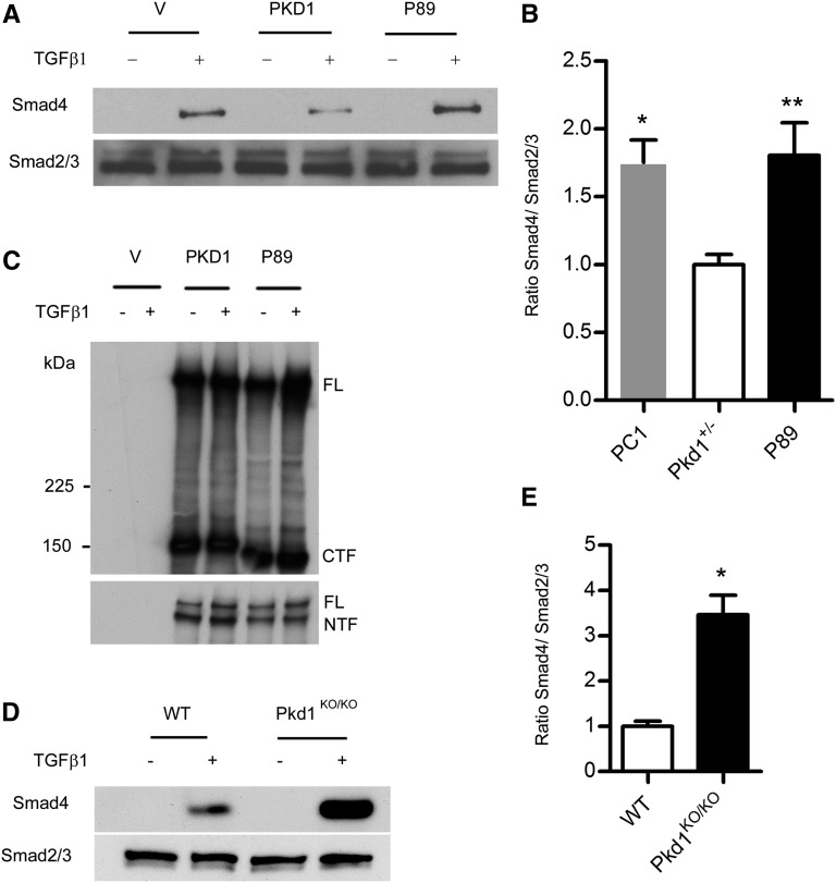
Pkd1 modulates responsiveness to TGF-β. (A–C) NIH3T3 cells are transiently transfected with flag-tagged human PKD1, flag-tagged P89 (a truncating PKD1 mutation, R4227×), or vector control (V) and then stimulated with 4 µg/ml TGF-β1. (A and B) Cell lysates are immunoprecipitated with an antibody to total Smad2/3, and then immunoblotted with anti-Smad4 (top). Membranes are stripped and reprobed with anti-Smad2/3 as a loading control (bottom). A representative blot is shown in A. Quantification (±SEM) in B shows that PKD1 expressing cells exhibit decreased recruitment of Smad4 to the Smad2/3 complex compared with vector control and P89 (n=3 independent experiments). *P=0.01; **P=0.03. (C) A portion of the cell lysate is immunoprecipitated with M2 beads and Western blots are probed with an antibody to the C-terminus (top) or N-terminus (bottom) of PC1. As expected, P89 is slightly smaller than wild-type PC1 but transfection efficiencies are comparable. (D and E) MEFs derived from Pkd1KO/KO or wild-type littermate controls are serum starved overnight and then incubated with 4 µg/ml TGF-β1. At baseline, there is little activation of canonical TGF-β signaling. After stimulation with TGF-β1, however, Pkd1KO/KO MEFs exhibit increased recruitment of Smad4 to the Smad2/3 complex compared with controls. (E) Quantification (±SEM) of three experiments. *P<0.01. PC1, polycystin-1; FL, full-length PC1; CTF, C-terminal cleavage fragment; NTF, N-terminal cleavage fragment.

