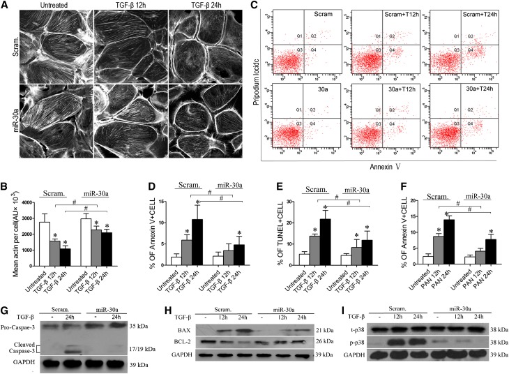
Figure 3.
Exogenous miR-30a protects podocytes from cytoskeletal damage and apoptosis induced by TGF-β or PAN. (A) Fluorescein-conjugated phalloidin staining of stably transfected Scram or miR-30a podocytes after treatment with TGF-β (5 ng/ml). (B) Quantification of results in A. (C) Annexin V flow cytometry of Scram or miR-30a podocytes after TGF-β treatment for 12 or 24 hours. (D) Quantification of results in C. (E) TUNEL assay of the cells as in C. Each bar represents the mean ± SD of the percentages of apoptotic cells in 30 high-power fields. (F) Annexin V flow cytometry of Scram or miR-30a podocytes after PAN treatment (50 µg/ml) for 12 or 24 hours. (G–I) Immunoblotting of cleaved caspase 3 (G), BAX and BCL2 (H), and p38 (t-p38) and phosphorylated p38 (p-p38) (I) in the Scram and miR-30a podocytes after TGF-β treatment for the indicated time. All data are presented as the mean ± SD of three independent experiments. *P<0.05 versus untreated control; #P<0.05.

