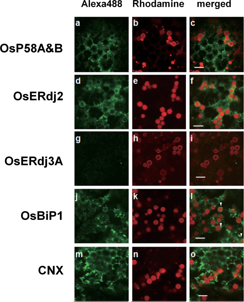
Fig. 6.
Subcellular distribution of OsP58A&B, OsERdj2, and OsERdj3A in wild-type seeds. Left panels show localization of OsP58A&B (a), OsERdj2 (d), OsERdj3A (g), OsBiP1 (j), and calnexin (CNX) (m); middle panels (b, e, h, k, and n) show localization of PB-Is (red); and right panels (c, f, i, l, and o) show the merged images of the left and middle panels. Arrowheads in l indicate PB-Is covered with OsBiP1 antibody. CNX was employed as a representative ER-marker protein. Bar=5 µm.

