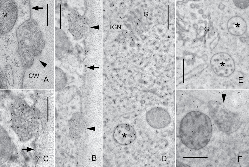
Fig. 9.
Immunodetection of the ARA6-related protein on EM sections of internodal cells of C. braunii. (A) Unstained longitudinal section. Smooth plasma membrane (arrow) and a charasome (convoluted plasma membrane; arrow head) are seen adjacent to the cell wall (CW). M, mitochondrion. (B–D) Immunogold labelling of ARA6. Accumulation of gold particles is seen in charasomes (arrow heads) and at smooth plasma membrane regions (arrows; C is an enlarged detail of B), in multivesicular endosomes (asterisks) and at the TGN. G, Golgi body. (E, F) Negative controls with pre-immune serum instead of primary antibody. A few immunogold particles are randomly distributed over the cytoplasm and the cell wall. Bars, 500nm.

