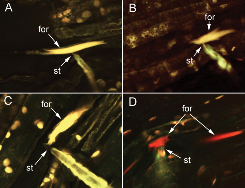
Fig. 3.
Confocal micrographs illustrating examples of forisomes (for) classified as (A) condensed; (B and C) intermediate; and (D) dispersed. Stylet tips (st) are in contact with the forisome in all 4 micrographs. Note in (D) that while the forisome in the penetrated sieve element is dispersed, a forisome in a neighbouring sieve element (right side of micrograph) is condensed. Samples were double stained with DiOC7(3) and sulphorhodamine 101 (Molecular Probes, Eugene, OR USA).

