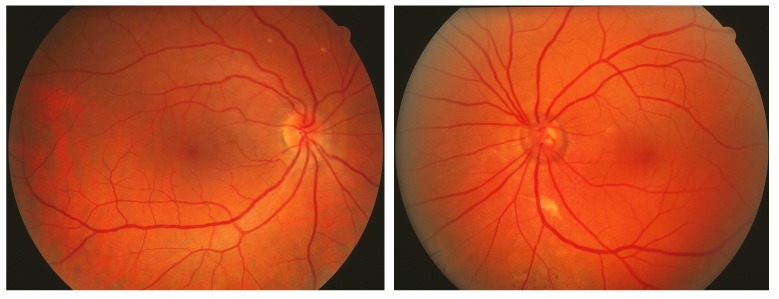
FIGURE 2.
Patient with port-wine stain involving the entire left side of face with left ocular fundus darker red, without normal choroidal vascular markings (patient 20, Table, also seen in Figures 10 and 16). Left, Normal right ocular fundus. Right, The left ocular fundus has a deeper red appearance due to the thickened blood-filled choroidal layer. Expansion of the choriocapillaris often dissimulates underlying choroidal channels and details otherwise visible. Such subtle changes, which give rise to the so-called tomato-catsup fundus, are best noted when there is a normal contralateral eye (left photo) for comparison and are often overlooked whenever venous hypertension is present bilaterally. The cup-disc ratio is increased in the left eye, indicative of the patient’s glaucoma with nasal visual field loss.

