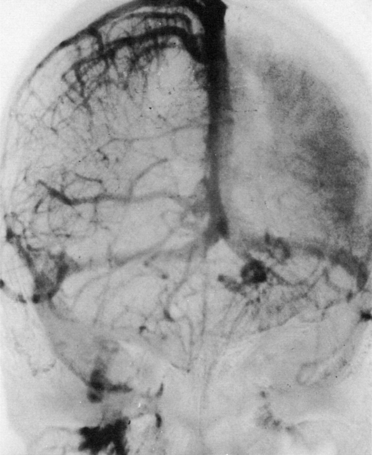
FIGURE 4.
Bilateral simultaneous internal carotid artery injection angiogram, mid-venous phase, anteroposterior projection in patient with Sturge-Weber syndrome. An 8-year-old girl with port-wine stain affecting the left forehead, right facial seizures, and right hemiparesis. The right superficial cerebral veins and the superior sagittal sinus are well filled. A striking lack of cortical veins over the left hemisphere can be noted. A prominent “brain stain” of the left cerebral hemisphere is related to delayed venous drainage. Enlargement of the internal cerebral and basal veins and their tributaries is evident. (Reprinted with permission from the Radiological Society of North America.2)

