Fig. 4.
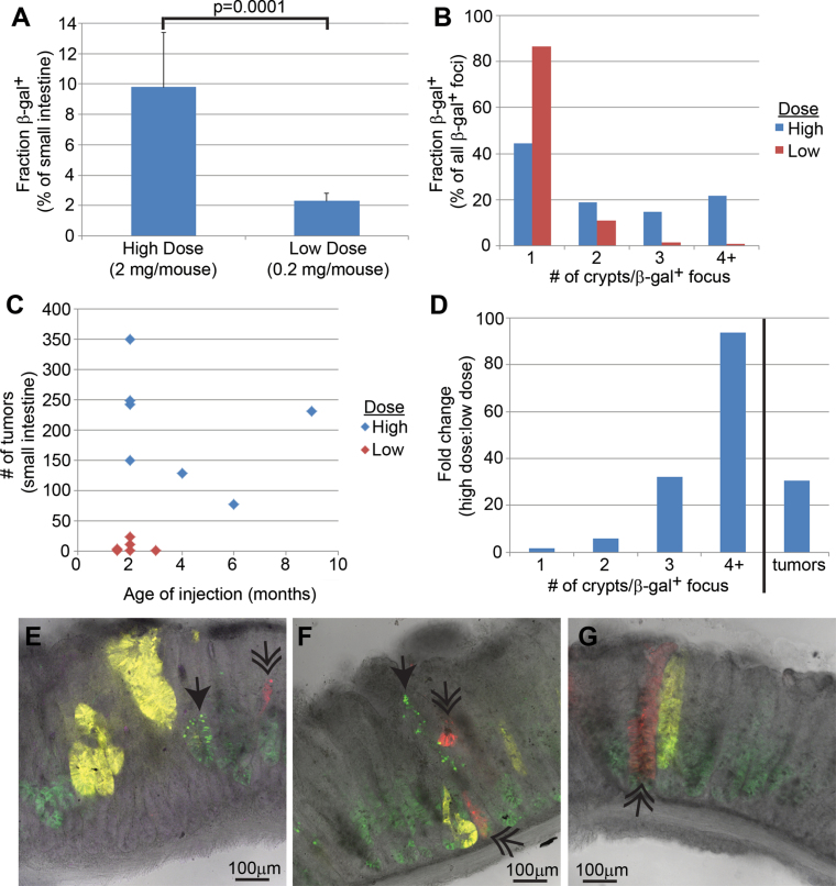
Characterization of recombination in Lgr5-CreER mice. (A) A 4-fold difference between the high (n = 4) and low (n = 8) dose of tamoxifen in the percentage of tissue with marker gene recombination. (B) The increased β-gal+ focus size in the intestine following high-dose tamoxifen as measured by the number of neighboring β-gal+ crypts. (C) The increased number of tumors in Lgr5-CreER; Apc CKO/CKO intestine following high-dose tamoxifen. (D) The higher dose of tamoxifen gives a higher ratio of larger β-gal+ fields. As discussed in the text, we estimate that at least three neighboring crypts are required for efficient adenoma initiation. (E– G) Images of adenomas from an Lgr5-CreER; Apc 580S/580S; R26R-Confetti mouse injected with a single, high dose of tamoxifen. Notice the multiple different fluorescent proteins found in single adenomas [nuclear green fluorescent protein (single arrowhead), red fluorescent protein (double arrowhead) and yellow fluorescent protein]. Cytoplasmic green fluorescent protein is from the Lgr5-targeted transgene.

