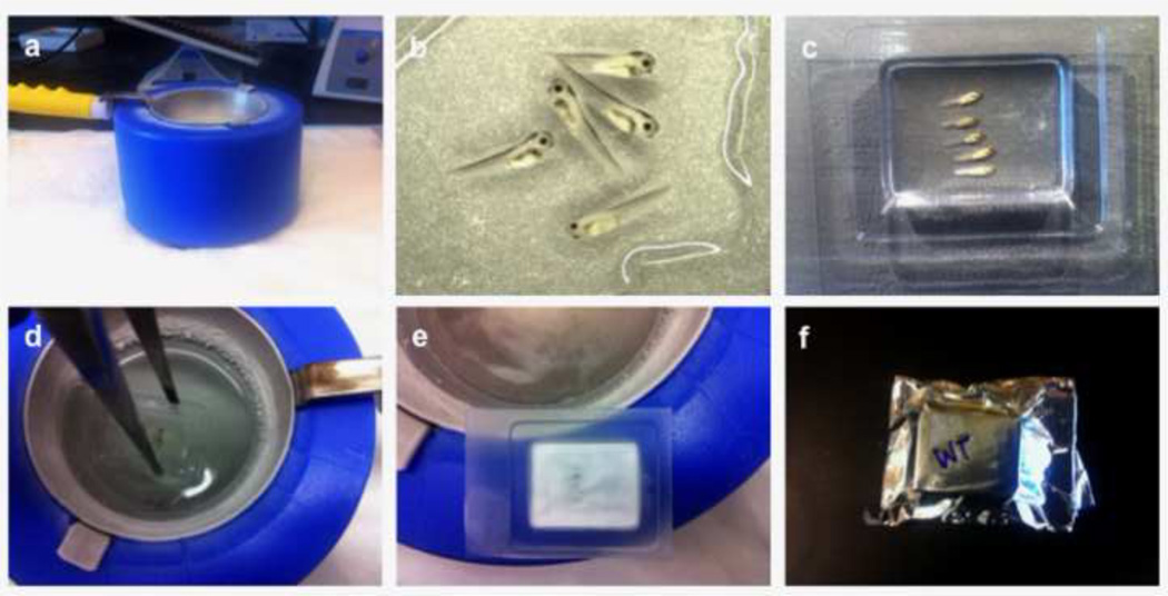
Figure 2. Cryosection preparation.
A liquid nitrogen bath setup with an isopentane filled ladle (a). 4–5 embryos are cleared of all residual sucrose solution and covered with OCT freezing medium (b). The embryos are transferred to, and aligned in a cryomold (c) and then immersed into the isopentane bath (d) until they turn opaque (e). Samples are quickly allowed to dry, wrapped in aluminum foil and stored (e, f).

