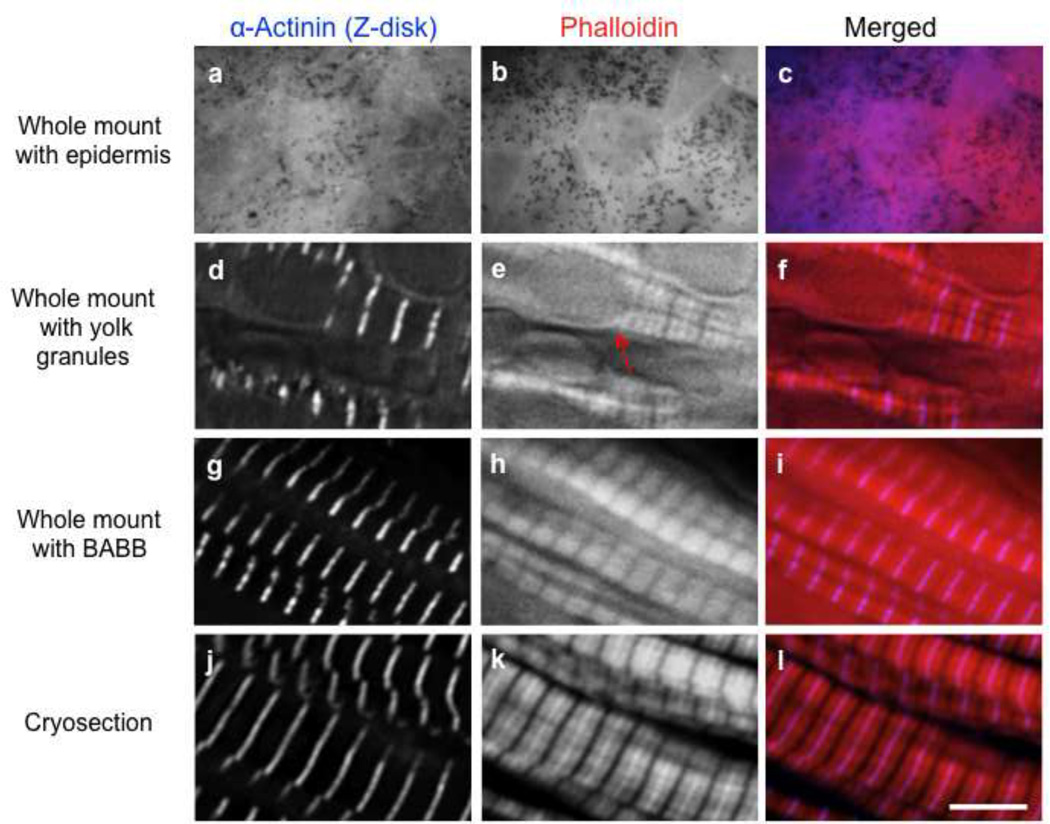
Figure 5. Comparison of stains from whole-mount, cryosection and BABB approaches.
When the epidermis is left on the whole-mount tissue, it obstructs the observation of myofibrils/sarcomeric proteins (a–c). Whole-mounts free of the epidermis exhibit staining for sarcomeric proteins (d–f) but yolk granules remain visible and obstruct subcellular structures (e, arrow). After staining, clearing somitic tissue with BABB masks yolk platelets; phalloidin labeling is preserved (g–i). Cryosections are superior for staining of actin filaments and sarcomeric proteins (j–l). Scale bar is 5 µm.

