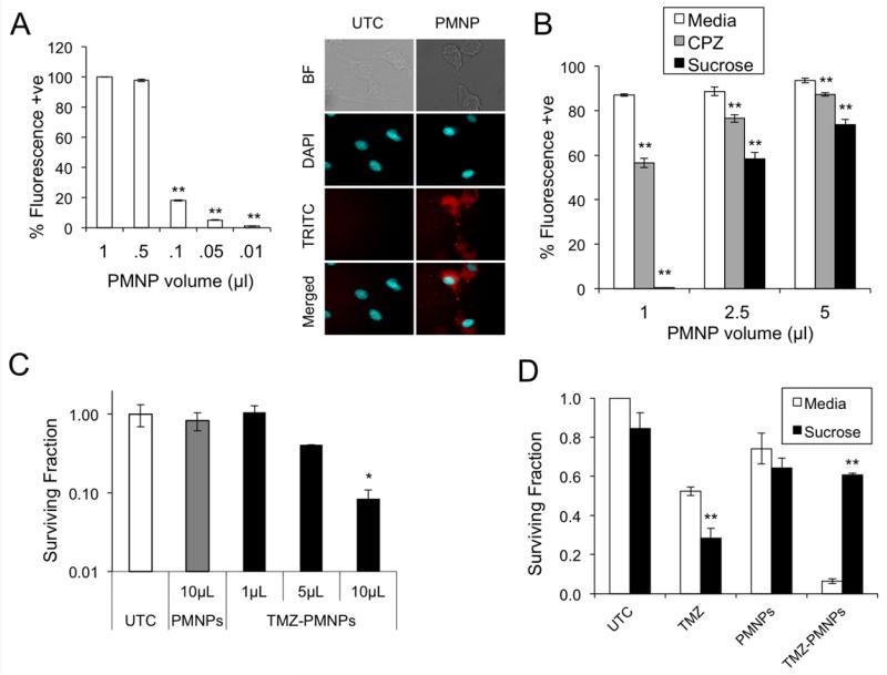
Figure 4. PMNPs are taken up by glioma cells and deliver viable TMZ.
(A) Uptake of PMNPs by U87 cells as demonstrated by FACS analysis (left) and confocal imaging (right) following exposure to rhodamine-tagged PMNPs (NP-Rhod) or rhodamine dye alone (UTC) for 2 hours. Rhodamine (TRITC) and nuclei (DAPI) were imaged as shown. (B) FACS analysis of PMNP uptake in the presence of the endocytosis inhibitors, chlorpromazine (30 μM) or hypertonic sucrose (450 mM). (C) Clonogenic assay of U87 cells following treatment with increasing amounts of TMZ loaded PMNPs (TMZ NP) or blank PMNPs. (D) Clonogenic assay of U87 cells, in the presence of 450 mM sucrose or regular media, following treatment with TMZ (50 μM), blank PMNPs, or TMZ-carrying PMNPs (carrying 50 μM TMZ). Clonogenic data show mean surviving fraction, normalized to untreated, of triplicate samples +/− SD. * P < 0.05 and ** P < 0.01 relative to untreated control.

