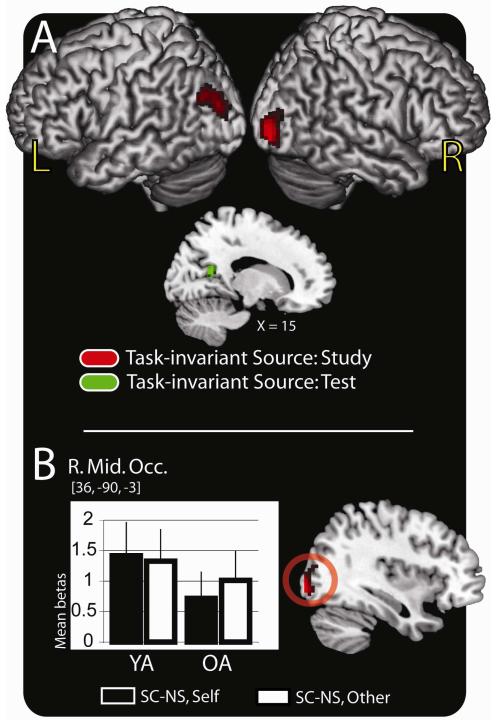
Figure 3.
Task-invariant activity at study and test. (A) Source memory regions (SC > NS) exhibiting task-invariant activation common to both younger and older adults at the time of study (red colors) and at the time of test (green colors) are rendered on a standard brain in MNI space. (B) Anatomic overlays and graphs depict mean parameter estimates (betas) from the peak voxel exhibiting task-invariant activation at the time of study. Bars represent, from left to right, the difference between the SC and NS trials in the self-reference task and the difference between the SC and NS trials in the other-reference task in the young adults (YA) followed by the same values in the older adults (OA). Bars are plotted in arbitrary units with the error bars depicting the SEM. Statistical threshold: p < .001, 34 contiguous voxels, corrected for multiple comparisons. MNI = Montreal Neurological Institute; SC = source correct; NS = no source; R. Mid. Occ. = right middle occipital cortex; SEM = standard error of the mean

