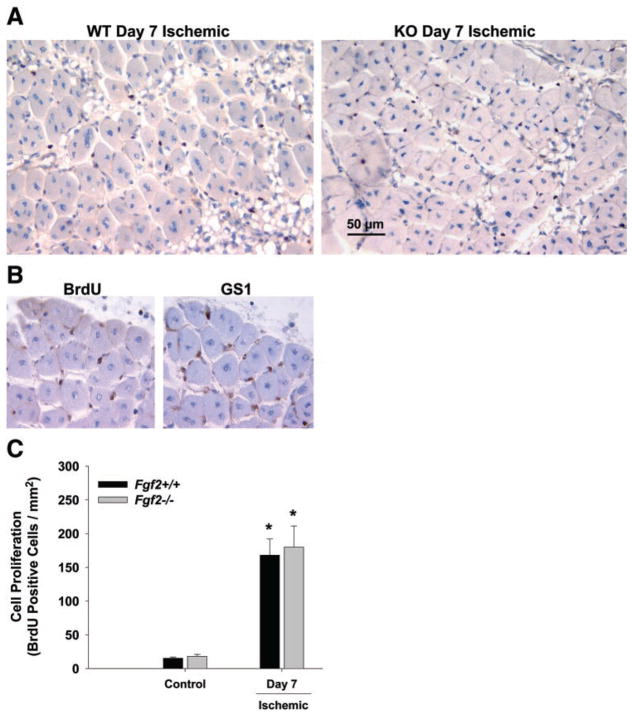
Fig. 5.
A: representative photomicrographs of muscle cross sections from day 7 postischemic hindlimbs of Fgf 2+/+ (WT) and Fgf 2−/− (KO) that are stained with bromodeoxyuridine (BrdU; blackish-brown stained nuclei) to identify proliferating cells. B: serial sections (6 μm each) from hindlimb stained with BrdU or GS1 to identify proliferating endothelial cells. C: quantification of cell proliferation in hindlimb muscle without femoral ligation (control) and 7 days after ligation.*P < 0.05 vs. respective control. Fgf 2+/+ vs. Fgf 2−/−, not significantly different.

