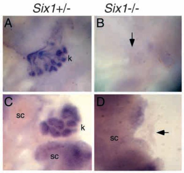
Fig. 7.
Six1 mutant mesenchyme is unresponsive to induction. (A) E11.0 Six1+/− kidney rudiments cultured for 5 days and stained with Pax2 in situ probe. After 5 days of culture, they developed into a fully branched kidney structure (k) showing Pax2 expression in the collecting tubules and in the nephrons. (B) E11.0 Six1−/− metanephric rudiments cultured for 5 days and stained with Pax2 in situ probe. No kidney formation was observed (arrow). (C) E11.0 Six1+/− metanephric mesenchyme cultured with spinal cord (sc) for 5 days and stained with Pax2 in situ probe. The Six1+/− mesenchymal cultures exhibited characteristic tubules (k) showing Pax2 expression. (D) E11.0 Six1−/− metanephric mesenchyme cultured with hetorozygous spinal cord for 5 days and stained with Pax2 in situ probe. None of the Six1−/− mesenchymes exhibited any sign of tubule formation. Note the disappearance of Six1 mutant mesenchyme (arrow), which shows no Pax2 expression.

