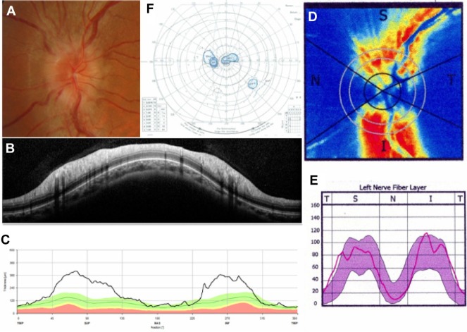
Figure 2.
Representative case of optic neuritis.
Notes: A fundus photograph (A) shows optic disc swelling in the left eye. Spectralis SD-OCT (B and C) and SLP with GDx ECC (D and E) reports demonstrate RNFL thickening. Goldmann perimetry (F) shows a central scotoma. Spectralis SD-OCT, Heidelberg Engineering Inc, Heidelberg, Germany; GDx ECC, Carl Zeiss Meditec AG, Jena, Germany.
Abbreviations: I, inferior; N, nasal; RNFL, retinal nerve fiber layer thickness; S, superior; SD-OCT, spectral-domain optical coherence tomography; T, temporal; SLP, scanning laser polarimetry.

