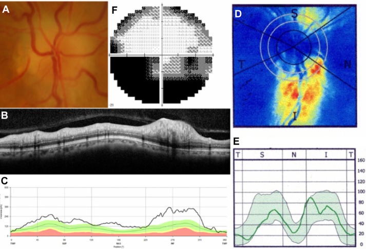
Figure 3.
Representative case of nonarteritic anterior ischemic optic neuropathy.
Notes: Fundus photograph (A) shows optic disc swelling with disc hemorrhage in the right eye. Spectralis SD-OCT images (B and C) show RNFL thickening in the superior quadrant, while SLP with GDx ECC imaging (D and E) shows slight RNFL thinning in the superior quadrant. Visual field testing (F) shows an inferior altitudinal field defect. Spectralis SD-OCT, Heidelberg Engineering Inc, Heidelberg, Germany; GDx ECC, Carl Zeiss Meditec AG, Jena, Germany.
Abbreviations: I, inferior; N, nasal; RNFL, retinal nerve fiber layer; S, superior; SD-OCT, spectral-domain optical coherence tomography; T, temporal; SLP, scanning laser polarimetry.

