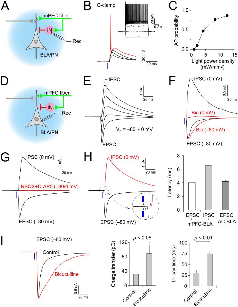
Figure 4.
Activation of mPFC Afferents Leads to Feed-Forward Inhibition of BLA Principal Neurons (A) Experimental design for recording of light-evoked (blue) synaptic responses in local circuit interneurons (IN) in the BLA. (B) BLA interneuron exhibits non-accommodating AP firing (inset). Synaptic responses were induced in BLA interneuron by photostimulation of mPFC projections in current clamp mode with stronger light intensities resulting in AP firing (red trace). (C) Probability of AP firing in BLA interneurons in response to photostimulation of the mPFC projections. It was assayed in cell-attached or current-clamp modes at resting membrane potential (n= 10 neurons). (D) Experimental design for recording of the light-evoked EPSC/IPSC in BLA principal neurons (BLA/PN). (E) The EPSC/IPSC in a BLA/PN at holding potentials ranging from −80 mV to 0 mV. (F) Synaptic currents recorded at 0 mV were completely blocked by GABAa receptor antagonist, bicuculline (30 μM, Bic; red trace at 0 mV). However, the peak amplitude of synaptic currents at −80 mV was not affected by bicuculline (red trace at −80 mV). (G) Both EPSC and IPSC (recorded at −80 mV or 0 mV, respectively) were blocked by jointly-applied NBQX (10 μM) and D-AP5 (50 μM) (red traces at −80 mV and 0 mV). (H) Left, synaptic latency analysis for EPSCs and IPSCs recorded in BLA/PN. Right, synaptic latency of IPSCs in the mPFC-BLA pathway (n = 19 cells) was significantly longer compared to photostimulation-induced EPSCs (n = 22 cells) or EPSCs induced by electrical stimulation of inputs to the BLA from the auditory cortex (AC, n = 21 cells). (I) Left, photostimulation-induced EPSCs in a BLA/PN (12.5 mW/mm2, 1-ms duration) under control conditions (black trace) and in the presence of bicuculline (30 μM, red trace). Right, average total charge transfer and 90%-to-10% decay time of EPSCs recorded in the presence or absence of bicuculline (n = 6 neurons from 4 mice). Error bars are SEM.

