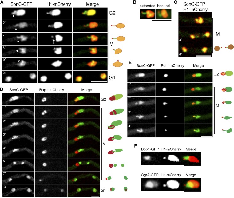
Figure 7.
SonC localizes to the NOR. (A) Time-lapse confocal microscopy of SonC-GFP in conjunction with histone H1-mCherry. Strain JLA319 (sonC-GFP H1-mCherry) was inoculated in minimal media and grown at room temperature for 24 h prior to imaging. The projection of SonC-GFP that corresponds to the NOR is marked by arrowheads. H1-mCherry is evicted from the middle region of the projection during mitotic chromosome condensation prior to anaphase (arrow) while SonC-GFP remains associated with the projection. Note that SonC-GFP does not locate to the distal H1-mCherry focus. Diagrams of the SonC and H1 localizations at the different time points are shown on the right. See corresponding File S6 and File S7. (B) The SonC projection can be visualized as an extended or a hooked structure. Strain is the same as in A. See the corresponding File S8 for hooked structure. (C) The SonC projection tipped by H1 was sometimes seen as lagging chromosome arms. Strain and growth conditions are the same as in A. See File S9. (D) Time-lapse confocal microscopy of SonC-GFP and the nucleolar marker Bop1-mCherry. The SonC-GFP projection is cradled within a portion of the nucleolar signal from Bop1-mCherry. Strain JLA324 (sonC-GFP bop1-mCherry) was treated as in A. Diagrams of the SonC and Bop1 localizations at different time points are shown on the right. See File S10 and File S11. (E) Time-lapse confocal microscopy of strain JLA325 (sonC-GFP pol I-mCherry). Strain was treated as in A. Diagrams of the SonC and Pol I localizations at different time points are shown on the right. See File S12. (F) The H1 focus is located outside of the nucleolus near the nuclear periphery. The position of the nucleolus is marked by the nucleolar proteins Bop1-GFP (strain LU193) or CgrA-GFP (strain LU178). The brightness and contrast of the images were adjusted using ImageJ software to highlight the relationship of the SonC projection with the other nuclear proteins more clearly. Bars, 5 μm.

