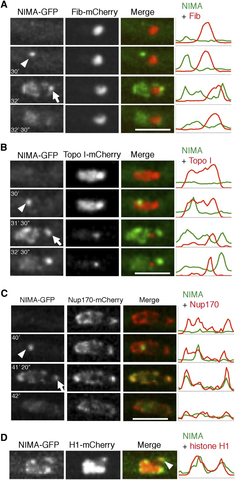
Figure 8.
NIMA locates to a NPC cluster adjacent to the nucleolus and the distal H1 focus during mitosis. (A) The location of NIMA-GFP in relation to the nucleolar marker Fib-mCherry was monitored in strain KF120 during mitosis. NIMA-GFP first locates to the SPBs (arrowhead), spreads around the nucleus, and locates to a second focus located on the nuclear envelope near the nucleolus (arrow). (B) The location of NIMA-GFP in relation to the NOR as defined by Topo I-mCherry was followed in strain KF122. After NIMA-GFP locates to the SPBs (arrowhead) during mitotic entry, it then locates to a second focus adjacent to the NOR as mitosis proceeds (arrow). (C) The localization of NIMA-GFP in relation to NPCs marked using Nup170-mCherry was followed in strain KF110. Only after initially locating to the SPBs (arrowhead) does NIMA-GFP then colocalize with a NPC cluster (arrow). (D) NIMA-GFP localization was followed in relation to the distal H1 focus in strain KF018. The second NIMA focus is located in the region of the nuclear envelope adjacent to the distal H1 focus (arrowhead). Pixel-intensity line profiles for A-D are shown to the right. Bars, 5 μm.

