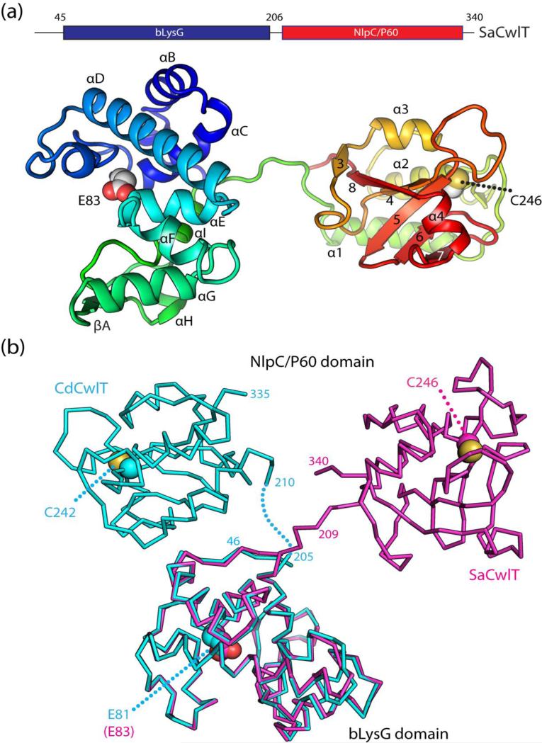
Fig. 3.
Crystal structures of SaCwlT and CdCwlT. (a) Ribbon representation of SaCwlT colored as a gradient from the N-terminus (blue) to the C-terminus (red). The secondary structure elements are labeled as defined in Fig. 2. Catalytic residues (Glu83: general acid and Cys246: nucleophile) are shown as spheres. (b) Comparison of SaCwlT (magenta) and CdCwlT (cyan). SaCwlT and CdCwlT are superimposed based on the bLysG domain.

