
Figure 2.
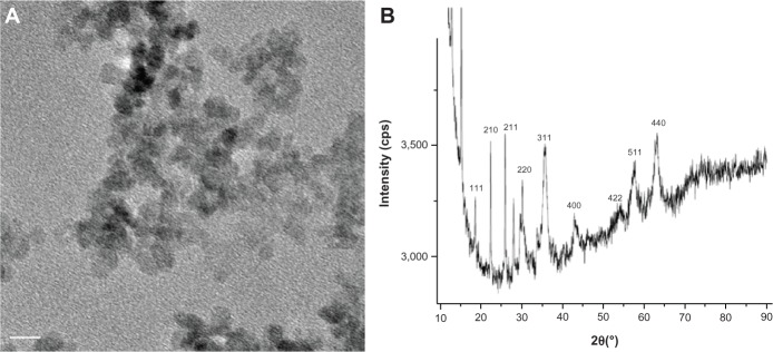
Transmission electron microscopy image and X-ray diffraction analysis of iron oxide nanoparticles coated with dextran.
Notes: (A) Transmission electron microscopy image and (B) X-ray diffraction analysis of iron oxide nanoparticles coated with dextran. The white bar corresponds to 10 nm.