
Figure 4.
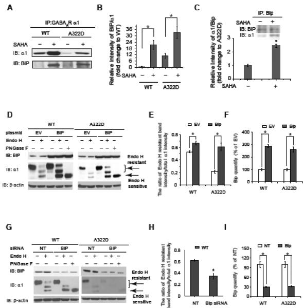
SAHA enhances the interaction between BiP and the α1 subunit, and BiP promotes the α1 subunit folding in the ER.
(A and B) SAHA treatment (2.5 μM, 24 h) enhances the interaction between the α1 subunit and BiP in HEK293 cells stably expressing WT α1β2γ2 or α1(A322D)β2γ2 GABAa receptors. Quantification of the relative intensity of BiP/α1 is shown in (B).
(C) Reverse immunoprecipitation confirms that SAHA treatment enhances the interaction between the α1(A322D) subunit and BiP.
(D, E and F) BiP overexpression increases endo H-resistant post-ER glycoform of the α1 subunit (n = 3) (D). PNGase F treatment serves as a control for unglycosylated α1 subunit. Quantification of the ratio of endo H-resistant / total α1 subunit bands is shown in (E), and quantification of BiP overexpression is shown in (F). EV: empty vector.
(G, H and I) BiP knockdown decreases endo H-resistant post-ER glycoform of the α1 subunit (n = 3) (G). Quantification of the ratio of endo H-resistant / total α1 subunit bands is shown in (H), and quantification of BiP knockdown is shown in (I). NT: non-targeting.
Each data point in (B), (C), (E), (F), (H) and (I) is reported as mean ± SEM. * p < 0.05