
Figure 1. The crystal structure of 14-3-3γ.
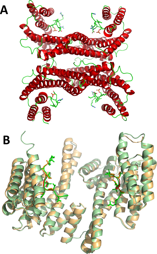
A) The two 14-3-3γ dimers (chains A–D) in the asymmetric unit (from this work; PDB 4J6S), as ribbons coloured by secondary structure, complexed to the TH peptides, represented by sticks coloured by atom type. B) A dimer of 14-3-3γ (chains A and B in PDB 4J6S), as pale green ribbons, with the TH peptides as bright green sticks, superimposed with the equivalent dimeric structure of 14-3-3γ from PDB 2B05, as pale orange ribbons, with the phosphopeptide (RAIpSLP) represented by red sticks.