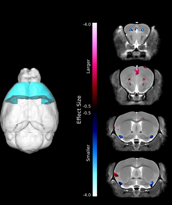
Figure 5.
(A) Highlighted is the frontal lobe, a region that commonly shows volumetric alterations in individuals with 22q11.2 microdeletions. B) Highlighted are the genotypic differences in relative volume within the frontal lobe region. Areas in red are significantly larger and areas in blue are significantly smaller in the Df(16)A+/− mice compared to WT littermates.

