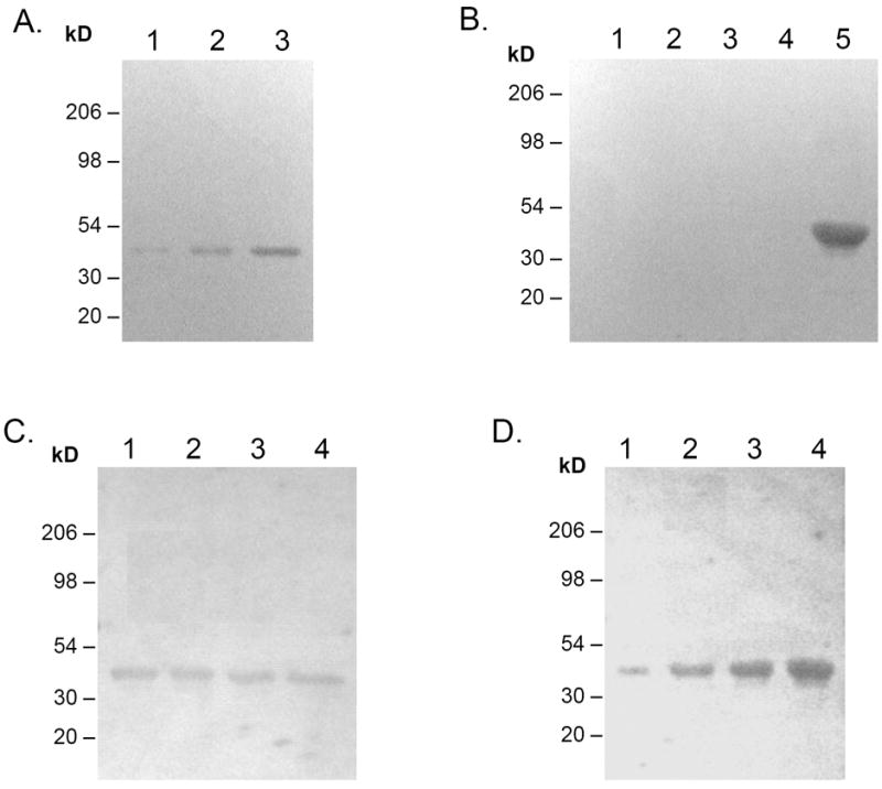
Figure 3. Characterization of antigen responsible for the observed antibody responses by western blot.

To determine antibody response to immunization, Pg extract or purified Pg fimbriae were blotted onto nitrocellulose as follows: (A) Various amount of Pg extracts was blotted onto nitrocellulose as follows: 1. 10μg, 2. 30μg, 3. 90μg. (B) Purified Pg components were blotted onto nitrocellulose as follows: 1. Lys-gingipain, 2. Gingipain R, 3. Gingipain R1, 4. Recombinant hemin/hemoglobin utilization receptor, 5. Purified Pg fimbriae (67kD + 41kD). (C) Lane 1-4, 100μg Pg extract each; (D) Lane 1-4, 1μg, 3μg, 6μg, 9μg of purified Pg fimbriae. The membrane was incubated with pooled serum (1:200) or pooled saliva (1:3.5) from rats immunized with Pg DNA (6 weeks post immunization), then with goat anti-rat IgG-HRP (1:10000). Color was developed with HRP substrate kit (BioRad).
