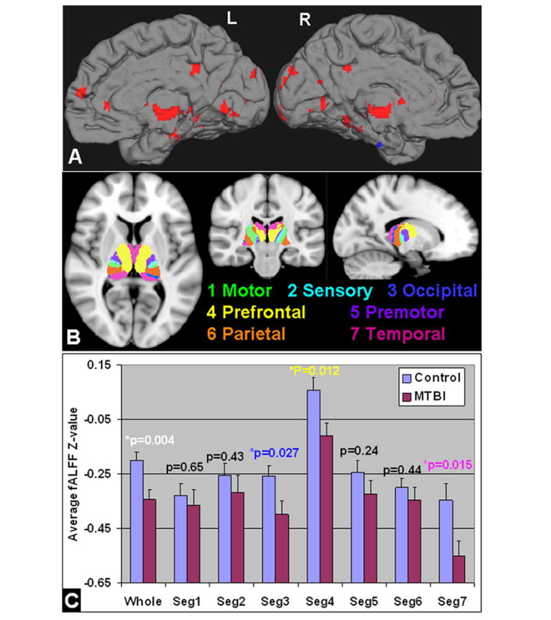
Figure 3.

A: fALFF analysis based on RS-fMRI comparing MTBI with controls in the whole brain showed mostly reduced fALFF in the thalamus, medial frontal, occipital and temporal regions (red color) (minimum Z>2.3; cluster significance, P<0.05, corrected). Results were showed in the medial surface projection views on the left and right hemispheres. Specifically, in the seven thalamic segments according to FSL template (B): there was significantly reduced fALFF in the overall thalamus and its segments for occipital (template index number 3), frontal (4) and temporal (7) projections in MTBI patients compared with controls (P<0.05) (C). As the fALFF was z-standardized over the whole brain; the negative values indicated the thalamic fALFF was less than the whole brain average.
