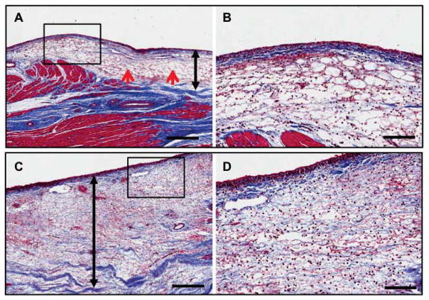
Figure 5.
Histopathological analyses after collagenase application: Representative images of trichrome elastic von Giessen staining of scar tissue after collagenase type 4 (CLG-4) application in (A) subject 1 and (C) subject 5 (scale bar, 250 μm). (B) and (D) indicate corresponding higher-power fields of (A) and (C), respectively (scale bar 100 μm). Red arrows indicate connective tissue loosening. Black double-sided arrows indicate the approximate range of digestion lesion.

