Figure 2.
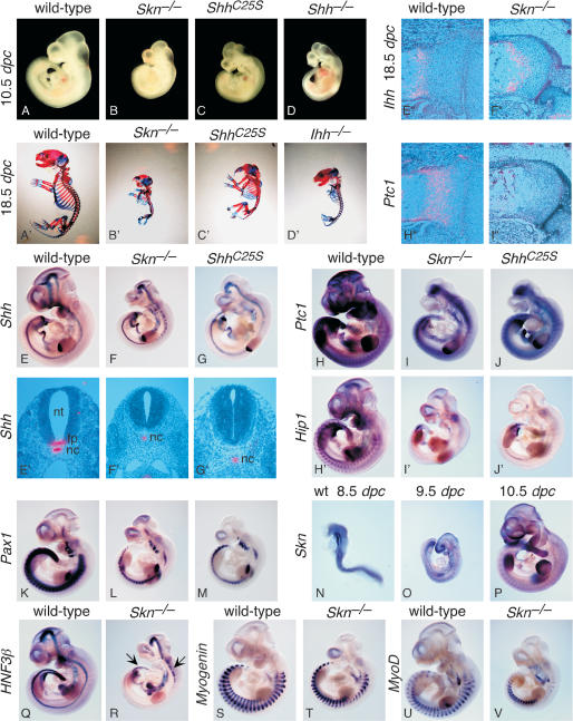
Skn and ShhC25S mutants exhibit multiple defects due to reduced Hh signaling. (A-D) External morphology of wild-type, Skn-/-, ShhC25S, and Shh-/- embryos at 10.5 dpc. All views are lateral. (A′-D′) Skeleton of wild-type, Skn-/-, ShhC25S, and Ihh-/- 18.5-dpc embryos stained with Alcian blue and Alizarin red. All views are lateral. Embryos in A-D and A′-D′ were photographed at the same magnification, respectively. (E-V,H′-J′) Whole-mount in situ hybridization, using digoxigenin-labeled riboprobes on wild-type, Skn-/-, and ShhC25S embryos at 10.5 dpc with the exceptions of N (8.5 dpc) and O (9.5 dpc). All views are lateral. Expression of Hip1 is known to be completely dependent on Hh signaling (Chuang and McMahon 1999), whereas Ptc1 expression is initially Hh independent but is strongly up-regulated upon Hh signal transduction (Goodrich et al. 1996). Arrows in R point to residual HNF3β expression in the more rostral and caudal part of the ventral midline of the neural tube. Expression of Myogenin and MyoD (Tajbakhsh et al. 1997) in Skn-/- mutants cannot be readily distinguished from that in wild-type embryos. Embryos in E-G; H-J; H′-J′; K-M; Q and R; S and T; and U and V were photographed at the same magnification, respectively. (E′-G′) Isotopic in situ hybridization using 33P-UTP-labeled riboprobes (pink) on paraffin sections of wild-type, Skn-/-, and ShhC25S 10.5-dpc embryos at the forelimb/heart level. (E″,F″,H″,I″) Isotopic in situ hybridization using 33P-UTP-labeled riboprobes (pink) on paraffin sections of proximal tibia of wild-type and femur of Skn-/- 18.5-dpc embryos. Expression of Ptc1 in the chondrocytes is regulated by Ihh and not Shh (St-Jacques et al. 1999). (Nt) Neural tube; (fp) floor plate; (nc) notochord.

