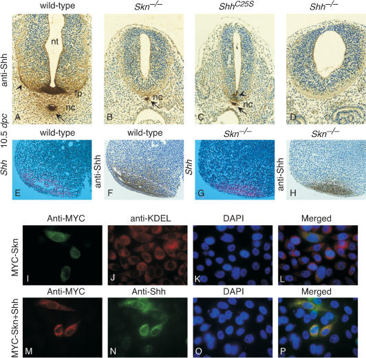
Figure 5.
Shh protein is mainly restricted to its sites of synthesis in Skn and ShhC25S mutants. (A-D) Cross-sections of wild-type, Skn-/-, ShhC25S, and Shh-/- embryos at 10.5 dpc at the forelimb/heart level stained with anti-Shh antibodies. In the wild-type section, Shh immunoreactivity (brown) is strong in the notochord and floor plate, and it extends out bidirectionally in a graded fashion (arrows and arrowheads). Similar patterns of Shh immunoreactivity extending from the notochord were observed on embryo sections where the floor plate has not yet been induced (Gritli-Linde et al. 2001), suggesting that Shh immunoreactivity in the neural tube is derived from both the notochord and floor plate. In sections of Skn-/- embryos, Shh immunoreactivity is mainly detected in the notochord (arrow), and no obvious extended staining is present. Similarly, Shh immunoreactivity is mainly detected in the notochord (arrow) of ShhC25S embryos. In this section (C), residual Shh immunoreactivity could be detected in the ventral midline of the neural tube (arrowhead), but no obvious extension of staining to the neural tube was noted. This is due to a more posterior extension (to the forelimb level) of residual Shh mRNA expression in the ventral midline of the ShhC25S neural tube at the more rostral levels (data not shown) than that in Skn mutants (Fig. 2R). Immunostaining was performed on multiple sections and on sections where Shh mRNA (Fig. 2E′-G′) or protein expression level in the notochord of Skn-/- and ShhC25S mutants was comparable to that of wild-type; lack of spreading of Shh immunoreactivity was still observed. No Shh immunoreactivity is detected in the notochord or neural tube of Shh-/- embryos, although Shh immunoreactivity is detected in the gut of Shh-/- embryos, which represents cross reactivity to Ihh epitopes (data not shown). (E,G) Isotopic in situ hybridization using 33P-UTP-labeled riboprobes (pink) on paraffin sections of wild-type and Skn-/- limbs at 10.5 dpc. F and H are adjacent sections to E and G and were stained with anti-Shh antibodies. The dotted pink lines in F and H represent the approximate domains of Shh mRNA expression as determined in E and G. Multiple limbs were sectioned, and in situ hybridization and immunostaining were performed on all sections from the limb. (I-L) CHO cells transiently transfected with an expression construct encoding a MYC-tagged Skn and stained with anti-MYC and anti-KDEL antibodies (ER marker). L is a merged image of I-K. (M-P) CHO cells cotransfected with expression constructs encoding MYC-SKN and Shh and stained with anti-MYC and anti-Shh antibodies. P is a merged image of M-O. Similar results were obtained by using HeLa cells (data not shown). (nt) Neural tube; (fp) floor plate; (nc) notochord.

