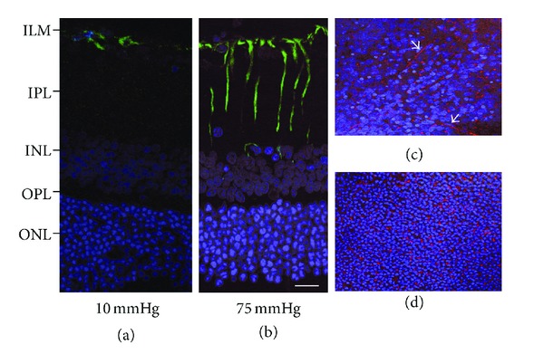
Figure 3.

Immunofluorescent localization of glial fibrillary acidic protein by confocal fluorescent microscopy. (a) GFAP expression is recognized as FITC fluorescence, and localized in the Müller cells endfeet of (arrow) in a normal retina (10 mmHg). (b) Apparent fluorescent reaction is recognized in the Müller cell body (arrowheads) and the Müller cells endfeet (arrow) at 75 mmHg. (c) and (d) Tangential view of the GFAP expression at the level of the inner nuclear layer (c) and the outer nuclear layer (d). GFAP-stained Müller cell bodies were recognized as (rhodamine-stained) reddish dots among the DAPI- (4′,6-diamidino-2-phenylindole dihydrochloride) stained nuclei. Note the obliquely running Müller cell process (arrows). Figures (a) to (d) are in the same magnification. Figures (a) to (d) are in the same magnification. Bar = 20 μm.
