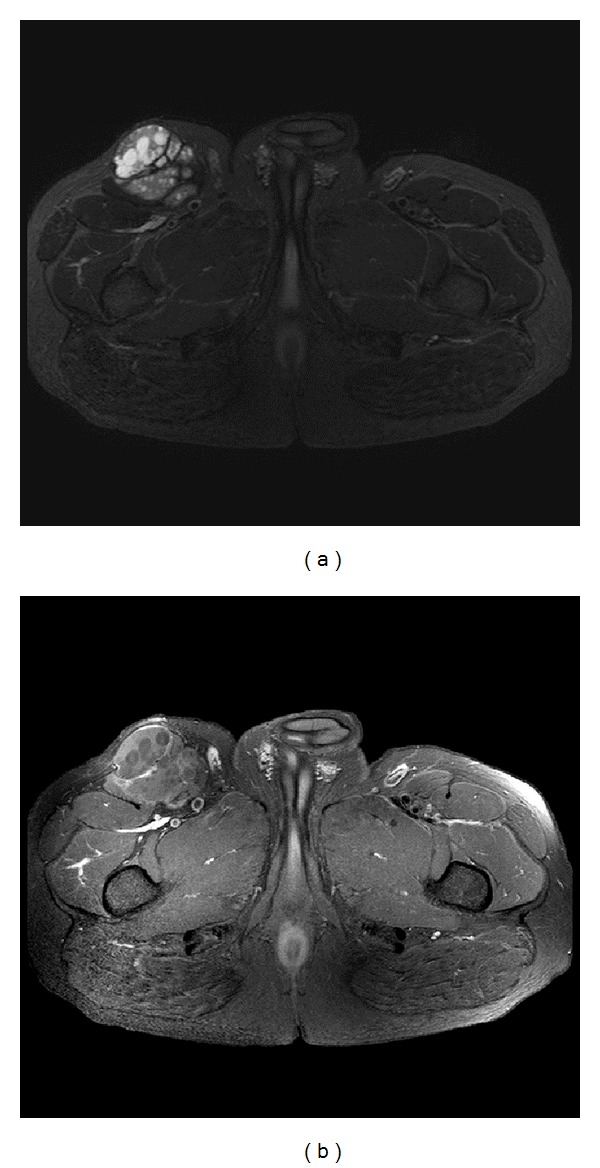
Figure 1.

(a) Axial T2 weighed image with fat saturation, it demonstrates a complex cystic lesion arising from the right sartorius muscle. It contains multiple small rounded daughter cysts and internal fibrous septae. No signs of invasion to the adjacent structures to suggest an aggressive sarcoma. (b) Axial T1 weighted image with fat saturation and postcontrast enhanced study: it demonstrates lack of enhancement of the complex lesion. The small rounded daughter cysts are nonenhancing consistent with hydatid cyst. Overall, the lack of enhancement as well as absence of aggressive features favors hydatid disease over soft tissue sarcoma.
