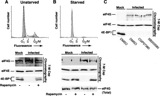
Figure 4.
Assembly of active eIF4F complexes in quiescent cells is enhanced following HSV-1 infection. Unstarved, asynchronous NHDF cells (A) and serum-starved, growth-arrested primary human NHDF cells (B) were either mock infected or infected with wild-type (WT) HSV-1 in the presence of DMSO or rapamycin. After 12 h, cell extracts were prepared and proteins bound to 7-methyl GTP Sepharose 4B were fractionated by SDS-PAGE, immunoblotted and visualized with the indicated antibodies. A sample of unfractionated extract prepared from starved cells was also analyzed by immunoblotting to determine the overall levels of eIF4G (eIF4G total). FACs profiles of uninfected serum-starved (72 h) or unstarved cells stained with propidium iodide appear above the immunoblots and peaks representing cells in various phases of the cell cycle (G1, S, G2/M) are indicated. (C) Serum-starved, growth-arrested NHDF cells were treated with either DMSO, the mnk-1 inhibitor CGP57380, or the p38 inhibitor SB203580. Extracts were chromatographed on 7-methyl GTP Sepharose and analyzed as described above in A and B.

