Figure 3.

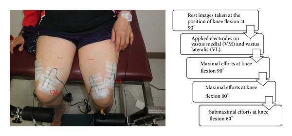
Examples of electrode positions for VM and VL placement. The ultrasound and s-EMG data is synchronously collected. The experimental protocol is listed, right.

Examples of electrode positions for VM and VL placement. The ultrasound and s-EMG data is synchronously collected. The experimental protocol is listed, right.