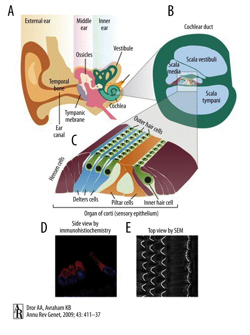
Figure 1.
Schematic illustration of the human ear. (A) The ear consists of the outer, middle, and inner ear. (B) A section through the cochlear duct illustrates the fluid-filled compartments of the inner ear. (C) The organ of Corti resides in the scala media, with sensory hair cells surrounded by supporting cells that include Deiters’, Hensen, and pillar cells. (D) Immunohistochemistry with the inner ear hair cell marker myosin VI, marking the cytoplasm of inner and outer hair cells, and4,6-diamidino-2-phenylindole (DAPI), marking the nuclei. (E) Scanning electron microscopy image of the top view of the sensory epithelium reveals the precise arrangement of 1 row of inner hair cells and 3 rows of outer hair cells, separated by the pillar cells. (with permission from Dror AA, Avraham KB. Hearing loss: Mechanisms revealed by genetics and cell biology. Annu Rev Genet, 2009; 43: 411–37. Copyright © 2009, Annual Reviews. All rights reserved).

