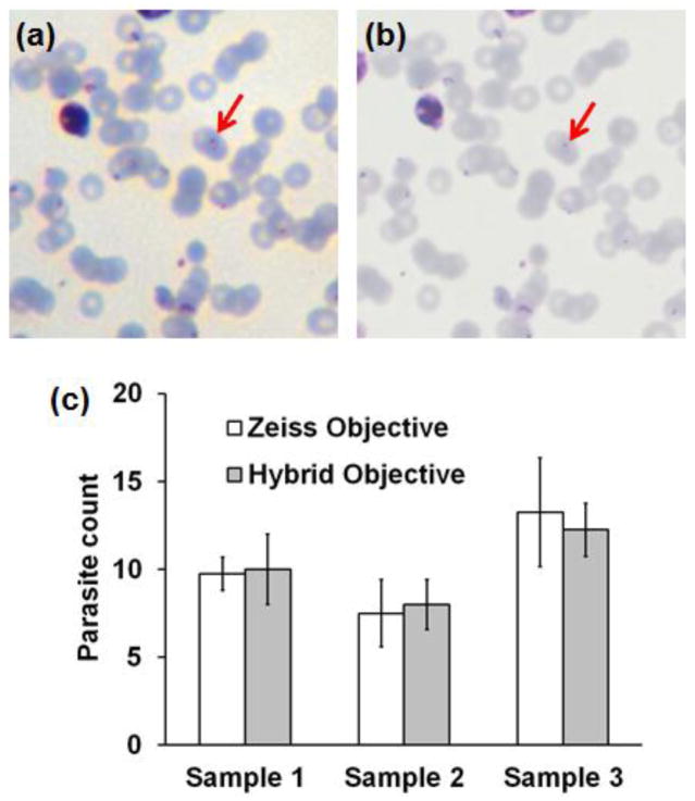
Figure 3.
Images of a malaria-infected human blood smear sample following Giemsa staining, acquired with (a) the hybrid objective, and (b) a Zeiss 40x / 0.95 plan-apochromat objective. (c) Comparison of parasite count determined in three paired fields-of-view, using images acquired by the hybrid and conventional objective systems.

