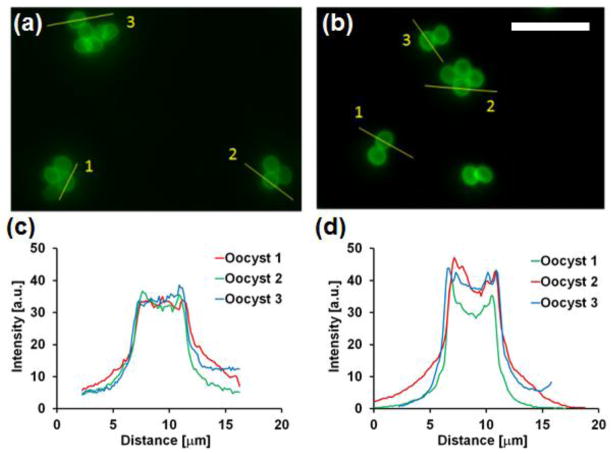
Figure 4.
Images of C. parvum immunofluorescent-stained oocysts taken with (a) the hybrid miniature objective and (b) a Zeiss 20x / 0.75 plan-apochromat objective. Scale bar = 20 m. Cross-sectional intensity profiles through individual oocysts from images acquired with (c) the hybrid and (d) conventional objective lenses.

