Figure 2.
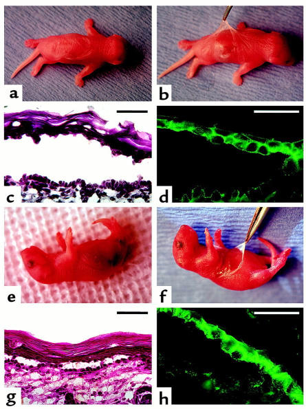
Induction of pemphigus in neonatal mice lacking Dsg 3 by passive transfer of IgGs from sera of PV patients that lack Dsg 1 antibody. Neonatal knockout Dsg3null mice homozygous for a targeted mutation of the Dsg3 gene (a–d) and “balding” Dsg3bal/Dsg3bal mice homozygous for a spontaneous null mutation in the Dsg3 gene (e–h) were injected intraperitoneally with 20 mg/g body weight of IgGs from the PV1 serum that lacked Dsg 1 antibody. Approximately 18–24 hours after injection, large, flaccid blisters filled with serous fluid were seen on the skin of both Dsg3null (a) and Dsg3bal/Dsg3bal (e) mice. At this time, the Nikolsky sign could be elicited on the skin of Dsg3null (b) and Dsg3bal/Dsg3bal (f) mice by mechanical extension of a large erosion after spontaneous rupture of the blister. The blisters developed due to intraepidermal separation showing suprabasilar acantholysis and prominent tombstone appearance of the basal cell layer (c and g; hematoxylin and eosin [H&E]). DIF with FITC-conjugated anti-human IgG antibody revealed intercellular staining of epidermis due to deposition of injected PV IgGs (d and h). All neonates of the progeny from mating of homozygous Dsg3null mice showed the PCR product of 280 bp, representing the sequence of the neomycin resistance gene used for the targeted disruption of the Dsg3 gene (17), and no 500-bp product, representing normal Dsg3 gene amplified from DNA extracted from a normal, Dsg 3–positive mouse (positive control). Likewise, all pups in the progeny from mating homozygous Dsg3bal/Dsg3bal mice lacked Dsg 3, as illustrated by finding a nonfunctional homozygous mutational insertion, 2275insT, upon direct nucleotide sequencing of the PCR product representing a portion of exon 14 of the mouse Dsg3 gene (data not shown). Scale bars = 50 μm.

