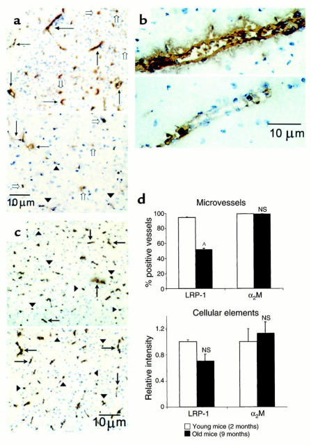
Figure 6.
(a) LRP-1 immunoreactivity in brain microvessels of young (2-month-old; upper panel) and old (9-month-old; lower panel) wild-type mice. Many vessels in young mice stained positive for LRP-1, detected with anti–LRP-1 Ab R777 (5 μg/ml; arrows). There were relatively fewer positive vessels in old mice (arrows), and many weakly positive- or negative-staining vessels (arrowheads). There was no significant difference in the staining of parenchymal cellular elements (open arrows) between the young and old mice. Vessels in young mice stained strongly positive (b, upper panel) compared with the faint staining seen in old mice (b, lower panel). In contrast, there was no difference in staining for α2M in brain cells (arrowheads) or microvessels (arrows) between young (c, upper panel) and old (c, lower panel) mice. (d) Comparison of LRP-1 and α2M immunoreactivity in brain microvessels (upper panel) and parenchymal cellular elements (lower panel) in young and old wild-type mice. AP < 0.05; NS, not significant.

