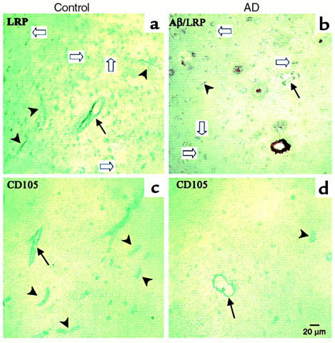
Figure 7.
LRP-1 expression in human frontal cortex. Brain sections (Brodmann’s area 10) of controls (a and c) reveal well-defined staining of capillaries (arrowheads) and arterioles (arrows) by LRP-1, detected with anti–LRP-1 mAb 8G1 (5 μg/ml) (a) and CD105 (c). No Aβ staining was present in double-labeled or serially labeled sections (not shown). In contrast, double-labeled sections from AD patients show vessels and plaque cores stained positive with anti-Aβ1-40 (brown stain), reduced numbers and intensity of LRP-1 staining of vessels (b), and reduced numbers of CD105-labeled vessels (d).

