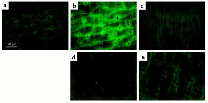
Figure 4.
ACh-induced production of H2O2 by the endothelium detected as an increase in fluorescence intensity in the DCF-loaded endothelial cells in small mesenteric arteries of mice. Fluorescence images of the endothelium in small mesenteric artery of a control mouse were obtained before (a) and 3 minutes after (b, d, and e) the application of ACh (10 μM). (c) Fluorescence image of smooth muscle layer of a control mouse obtained at the same visual field as a and b. The direction and depth of the smooth muscle layer were apparently different from those of the endothelial layer. (d) ACh-induced increase in fluorescence intensity was almost abolished by pretreatment with catalase (1250 U/ml) in a control mouse. (e) ACh-induced increase in the fluorescence intensity was markedly reduced in an eNOS-KO mouse. All experiments were performed in the presence of indomethacin (10 μM) and L-NNA (100 μM).

