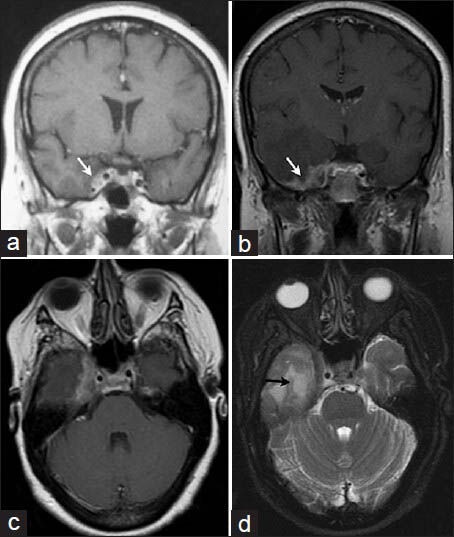
Figure 1.

First T1-weighted coronal magnetic resonance images (a) with contrast enhancement of a right cavernous sinus tumor (white arrow). Six months later, preoperative T1-weighted coronal (b) and axial (c) MRI with gadolinium showing progression of the tumor involving the right cavernous sinus with dura-arachnoid thickening and intense pachymeningeal enhancement with mesial temporal infiltration (white arrow); T2-weighted axial MRI (d) showing temporal lobe edema (black arrow)
