Figure 2.

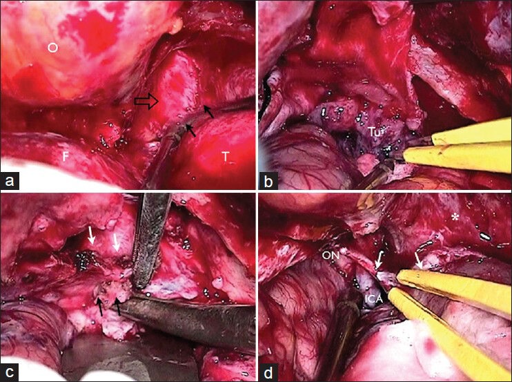
Intraoperative photos. (a) Right craneo-orbito-zygomatic with extended middle fossa approach, exposing the orbit (o), frontal (f), and temporal (t) lobes extradurally. Peeling of the lateral wall of the cavernous sinus and middle fossa floor identifying both dural layers (dura propia [black arrows] and dura periostica [open arrows]); (b and c) resection of tumor (Tu) with cavernous sinus and basal temporal dura propia infiltration (black arrows). Inner layer of cavernous sinus lateral wall without tumoral involvement (white arrows); (d) subtotal resection showing the optic nerve (ON), internal carotid artery (ICA), and dura mater (white arrows)
