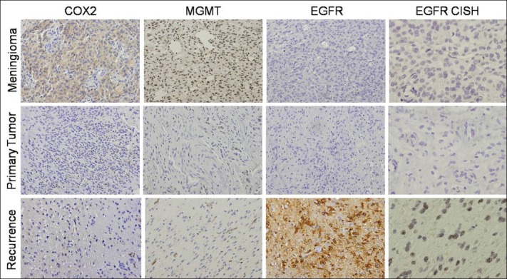
Figure 3.

Imunohistochemistry and Chromogenic In Situ Hybridization (CISH) analysis of the three lesions. COX2 immunohistochemistry (×200) positive for the meningioma lesion and negative for the primary and recurrent gliosarcoma. MGMT staining (×200) was only positive for the meningioma. EGFR immunostaining was negative in primary gliosarcoma and meningioma, with recurrent gliosarcoma exhibiting strong positivity. CISH analysis of EGFR confirmed these findings after EGFR amplification
