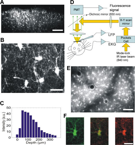
Figure 1. In Vivo Loading and Imaging of Astrocytes Using Fluo-4 AM.
(A) Acute slice prepared 1 h after dye loading. Scale bar, 200 μm.
(B) Higher magnification reveals cells with typical astrocyte morphology. Scale bar, 20 μm.
(C) Average bulk fluorescence as a function of the depth from the pial surface.
(D) Schematic drawing of the experimental arrangement. Abbreviations: EKG, electrocardiogram. PMT, photomultiplier. LFP, glass micropipe for local field potential and multiple unit recording. The same pipette was used to deliver bicuculline.
(E) Image taken 50–150 μm below pial surface in vivo. Flattened xyz stack.
(F) Fluo-4 AM loaded cells (left) were stained for S100B immunoreactivity (right), and the images were merged (center). See Video S3 for large-scale staining. Scale bar, 20 μm.

