
Figure 1. Ageing reduces ability to maintain SR Ca2+ content by increasing RyR-mediated Ca2+ leak.
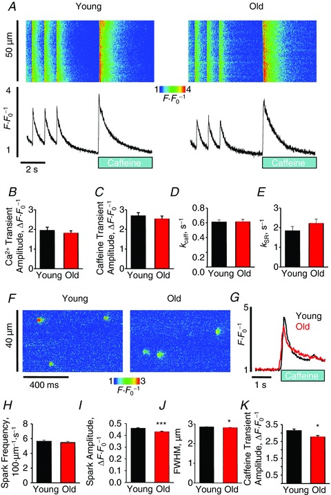
A, representative confocal line scan images and Fluo-3 F F0−1 profiles at 1 Hz. Graphs B–E depict mean data from Ca2+ transient amplitude (young, 1.94 ± 0.17 vs. old, 1.80 ± 0.13 ΔF F0−1), caffeine transient amplitude (2.69 ± 0.16 vs. 2.52 ± 0.16 ΔF F0−1), and decay rate constants (from exponential fit): kcaff: 0.61 ± 0.03 vs. 0.61 ± 0.04 s−1 and kSR: 1.83 ± 0.23 vs. 2.21 ± 0.23 s−1. For baseline Ca2+ transient, n = 12–23 cells from 3–4 heart preparations; all values P= NS and ± SEM. F, representative confocal line scan images with Fluo-4 of spark activity in saponin-permeabilized cardiomyocytes from young and old rabbits. G, representative Fluo-4 ΔF F0−1 profiles of caffeine-induced Ca2+ transients in permeabilized cardiomyocytes from young and old rabbits. Bar graphs H–K show mean data for spark frequency (young, (5.62 ± 0.18) × 100 μm−1 s−1 vs. old, (5.47 ± 0.17) × 100 μm−1 s−1, P = NS), spark amplitude (0.46 ± 0.004 vs. 0.43 ± 0.004 ΔF F0−1, ***P < 0.001), FWHM (2.85 ± 0.01 vs. 2.81 ± 0.01 μm, *P < 0.05), and caffeine transient amplitude (2.13 ± 0.10 vs. 1.76 ± 0.09 ΔF F0−1, *P < 0.05), respectively. For caffeine application, n = 20–35 cells, and for spark analysis n= 205–247 cells; n= 3431–4395 sparks from 3–4 heart preparations. All values are mean ± SEM.