
Figure 5. Oxygen sensitivity of TASK channels in type-1 cells.
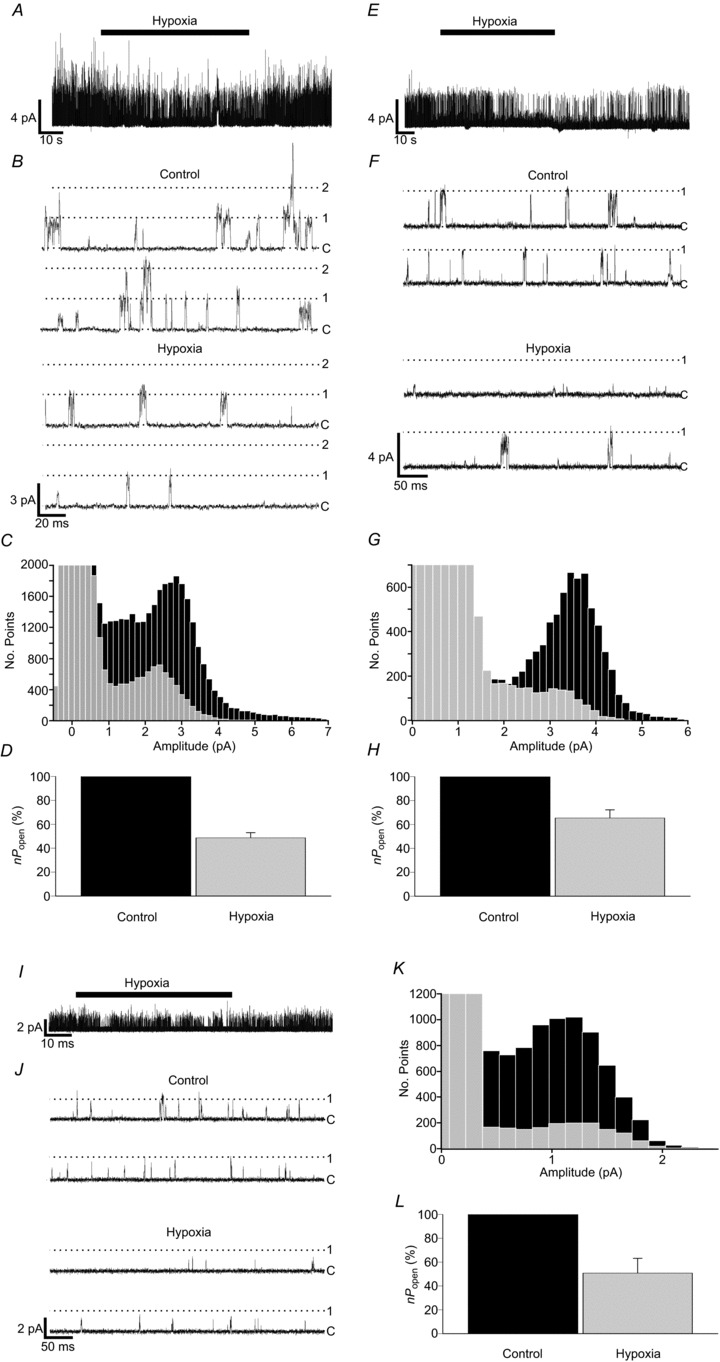
A, B, representative single-channel recording showing reversible inhibition of background channel activity by hypoxia in control type-1 cells. Cells were bathed in 100 mm K+, nominally Ca2+-free Tyrode solution. Hypoxia was induced by gassing Tyrode solutions with 5% CO2 and 95% N2. Pipette solution contained 140 mm K+, with a pipette potential of +80 mV. C, superimposition of all-points histograms derived from recordings made under control and hypoxic conditions. D, comparison of background channel activity under control and hypoxic conditions (n= 24 patches). E, F, single-channel recording from a Task-1−/− type-1 cell showing the reversibility of inhibition of channel activity by hypoxia. G, superimposed histograms constructed from Task-1−/− data obtained under control and hypoxic conditions. H, relative channel activity for Task-1−/− under control and hypoxic conditions (n= 16 patches). I, J, Task-3−/− single-channel activity inhibition by hypoxia. K, superimposition of all-points histograms created from Task-3−/− type-1 cell control and hypoxia recordings. L, comparison of relative channel activity in Task-3−/− cells under control and hypoxic conditions (n= 9 patches).