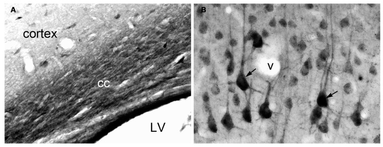
FIGURE 1.
Immunohistochemistry of NAA in specially fixed rat brain tissue using highly purified polyclonal antibodies to protein-coupled NAA. NAA immunoreactivity was observed in virtually all fiber pathways, such as the corpus callosum [cc in (A)]. In gray matter, NAA immunoreactivity was strong in large cortical pyramidal neurons [arrows in (B)], and moderate in smaller cortical neurons (B). NAA was also observed in many neuronal dendrites. Methods described in Moffett and Namboodiri (1995). Images taken with 20× (A) and 40× (B) objectives. LV, lateral ventricle; v, blood vessel.

