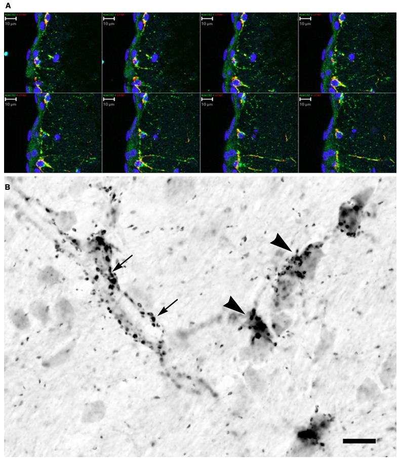
FIGURE 6.
AceCS2 immunoreactivity in the rat brain using an antibody generated against a 14 AA sequence from the C-terminus of murine AceCS2 (1:5000 dilution). Immunoreactivity in the adult rat brain was only observed in small punctuate structures, especially in white matter tracts, on some blood vessels and in the pia matter. (A) Confocal z-series images of immunofluorescence for AceCS2 (green) and the astrocyte marker GFAP (red) at the cortical surface including the pia matter (blue indicates DAPI staining of cell nuclei). Colocalization of AceCS2 and GFAP appears as yellow. This series of images show the merged images at various depths within the tissue slice. AceCS2 was associated with punctuate structures in astrocytes. (B) High magnification image of immunohistochemistry in adult rat corpus callosum. The punctuate structures were often closely apposed to blood vessels (arrows) or oligodendrocyte cell bodies (arrowheads). The stained structures ranged in size from ~½ to 1 micron. Bars in (A,B) = 10 μm.

