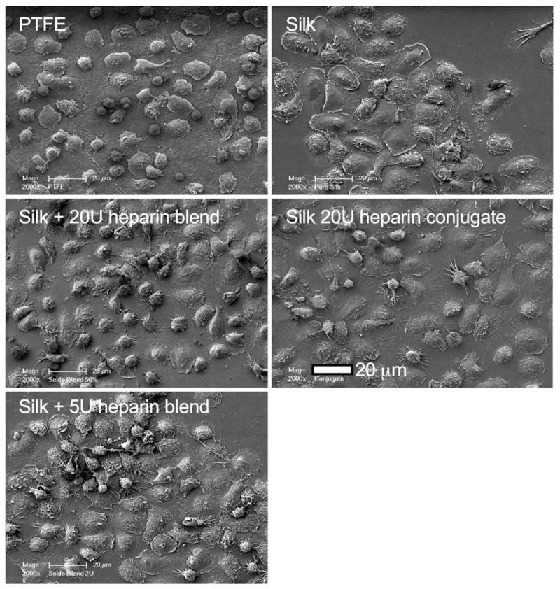
Figure 6.
Qualitative assessment of silk substrates exposed to whole human blood. Representative scanning electron micrographs of samples following a 2 h blood incubation. Images derived from PTFE co-incubation studies; comparable results were obtained for endothelial cell co-incubation studies. Samples are defined in Figure 4.

