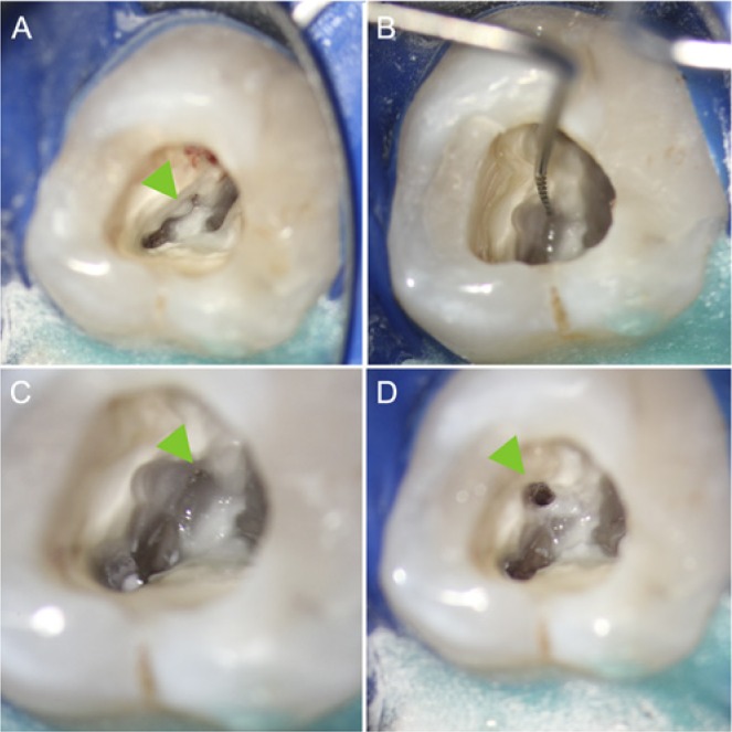
Figure 3.

Modern microendodontic procedure: identification and instrumentation of a calcified second mesiobuccal canal (MB2) in a first maxillary molar. (A) Overview after access cavity was prepared. Overhanging dentin covers MB2 (arrow)(10x magnification). (B) Identification of MB2 orifice with micro-instrument after ultrasonic removal of obstructing dentin (10x). (C) Initial preparation of MB2 (arrow) to allow for straight-line access (16x). (D) Fully instrumented MB2 canal prior to root filling (10x). Note mesial relocation of the orifice after complete debridement and instrumentation (arrow). Endodontic treatment by first author.
